From Shadow to Substance: The Arc of Imaging
To appreciate what drives Dr. Jobe, one must trace the remarkable evolution of medical imaging — a history he has not merely observed, but actively participated in and championed.
The era of plain radiography offered medicine its first systematic look beneath the skin. The plain X-ray was transformative in its time, casting bone into sharp relief against the ambiguous gray of soft tissue. Yet for a clinician focused on the tendons, ligaments, and nerves that animate the skeleton, the plain film was frustratingly mute. It showed the scaffolding but not the machinery. It revealed fracture but little of the inflammation, degeneration, or partial tear that so often explains a patient’s pain.
Computed tomography brought the first true revolution in anatomical detail, assembling cross-sectional slices into a three-dimensional portrait of the body’s interior. Bone pathology became far more legible, and complex joint architecture could at last be studied in depth. Yet soft tissue remained a secondary concern, and the technology demanded ionizing radiation and the confines of a large, fixed machine.
Magnetic resonance imaging arrived as a genuine wonder: rich, multi-planar images of soft tissue produced without radiation, capable of distinguishing a partial rotator cuff tear from a complete one, or identifying the edema within a ligament days after injury. Dr. Jobe recognized MRI as a powerful ally and has deployed it throughout his career. Yet he also came to understand its limitations — the static nature of the image, the expense, the inability to observe the body in motion, and the occasional ambiguity that sent clinician and patient alike back toward uncertainty.
And then came ultrasound…
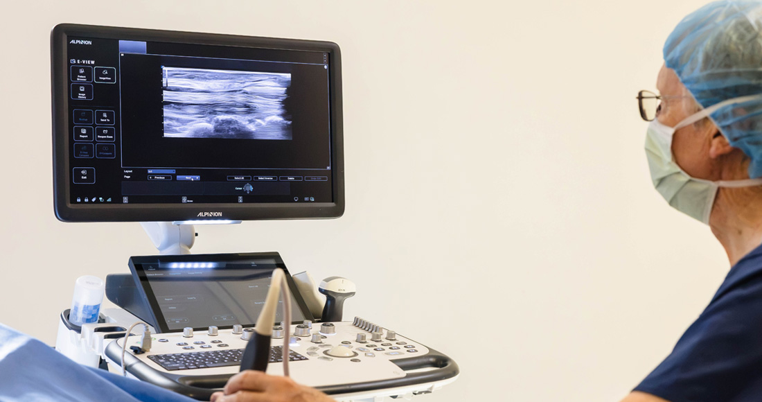
For Dr. Jobe, musculoskeletal ultrasound is not simply one more imaging tool — it is the culmination of the trajectory that began with Roentgen’s first X-ray films. Real-time, portable, radiation-free, and exquisitely sensitive to soft tissue pathology, ultrasound allows a skilled clinician to watch a tendon glide, observe a nerve compress, and assess a joint as the patient moves. It is, in the most literal sense, anatomy made dynamic. Dr. Jobe pursued and earned his RMSK certification in recognition of this conviction: that mastery of musculoskeletal sonography represents the current pinnacle of non-invasive diagnostic capability for the practicing clinician.
Diagnosis as the Foundation of Care
The evolution of imaging has always meant, for Dr. Jobe, an evolution in the precision of diagnosis — and precision in diagnosis is the precondition of precision in treatment. He has written and lectured on the clinical urgency of identifying the exact nature of an injury: not merely that a structure is damaged, but how, where, and to what degree. A partial-thickness rotator cuff tear, correctly identified on ultrasound, calls for a different therapeutic strategy than a full-thickness tear visualized on MRI. A compressed median nerve at the wrist, evaluated dynamically under ultrasound, may reveal surgical scarring invisible on static imaging. The right image, in the right hands, changes the clinical calculus entirely.
This commitment to diagnostic exactitude has driven Dr. Jobe’s academic work, including peer-reviewed case reports exploring the complementary roles of ultrasound and MRI in identifying pathology that neither modality fully captures alone. His clinical reporting has described the use of ultrasound-guided revision surgery in carpal tunnel release, relying on precise anatomical landmarks to navigate safely and effectively where prior intervention had left ambiguity and persistent symptoms. In each case, the lesson is the same: better imaging enables better outcomes.
The Biological Frontier: When Imaging Meets Regeneration
If advanced imaging represents one axis of Dr. Jobe’s enthusiasm, biological therapeutics represent the other — and the intersection of the two is where he finds the greatest excitement for medicine’s future.
The past two decades have witnessed the emergence of a new pharmacopeia drawn not from chemistry but from biology itself. Platelet-rich plasma (PRP) concentrates the growth factors naturally present in the blood, delivering them in high concentrations to sites of injury where normal healing has stalled. Bone marrow aspirate concentrate (BMAC) carries mesenchymal stem cells with the potential to differentiate into the tissues of the musculoskeletal system — cartilage, tendon, bone — in response to local biological signals. Extracellular vesicles and the broader microsecretome represent an even more refined horizon: the cell’s own signaling apparatus, harvested and applied therapeutically without the complexity of live-cell transfer.
Dr. Jobe has engaged deeply with this emerging science, both in reading the primary literature and in applying these modalities in practice. What captivates him is the synergy between biological treatment and precise imaging: to inject PRP or stem cells into a tendon tear with therapeutic intent requires knowing exactly where that tear is, how large it is, and what the surrounding tissue architecture looks like. Ultrasound-guided injection, performed in real time with needle visualization, transforms what would otherwise be an approximation into a targeted intervention. The biology and the imaging are, in this sense, inseparable. One without the other is incomplete.
This philosophy — that the value of a biological tool is only fully realized when deployed with imaging precision — is central to Dr. Jobe’s approach to regenerative medicine. He is not dazzled by the novelty of biologics for its own sake; he is excited by what becomes possible when the physician can see exactly where the therapy needs to go.
A Career Built on Curiosity
Beyond the clinical and academic dimensions of his work, Dr. Jobe is recognized by colleagues as a physician of broad intellectual formation. His engagement with the literature — across musculoskeletal ultrasound, regenerative medicine, peripheral nerve anatomy, and procedural technique — reflects a temperament that has never regarded medicine as a settled body of knowledge, but as a living discipline demanding continued inquiry.
He has lectured internationally, bringing his perspective on musculoskeletal diagnosis and ultrasound-guided technique to audiences beyond his own practice. He has contributed to the peer-reviewed literature through case reports that advance the field’s understanding of specific diagnostic and procedural challenges. And he has mentored in the tradition of a clinician who believes that knowledge, to be fully valuable, must be shared.
The story of Dr. Christopher M. Jobe is, in one sense, the story of medicine’s long march toward seeing more clearly and treating more precisely. From the ghostly silhouette of the first X-ray to the dynamic, real-time clarity of ultrasound; from the blunt instruments of inflammation management to the targeted biologics of regenerative medicine — Dr. Jobe has walked this arc with the enthusiasm of a scientist and the care of a healer. For him, every advance in imaging is not a destination, but an invitation: to look more closely, understand more fully, and treat more wisely.